cryo Diamond Knives
DiATOME cryo knives and their applications
- Thinner cryo sections
- Perfect cryosections from ultrathin to semi with the same knife
- Minimal compression and best structure preservation
- Highest quality diamonds and optimal crystal orientation guarantee perfect ultrathin sections and a durable cutting edge
| Knife type | Knife angle | Size [mm] |
Thickness range [nm] |
Boat type | Code (new knife)* |
Application |
|---|---|---|---|---|---|---|
| cryo 25° | 25° | 3.0mm |
30–150 | Triangular holder | 30-CD25 |
|
| cyro immuno | 35° | 2.0mm 3.0mm |
30–300 | Triangular holder | DCIMM3520 DCIMM3530 |
|
| cryo 35° (dry) | 35° |
1.5mm |
30–300 | Triangular holder | 15-CDL 20-CDL 25-CDL 30-CDL 35-CDL 40-CDL |
|
| cryo 35° (wet) | 35° | 1.5mm 2.0mm 2.5 mm 3.0 mm 3.5 mm 4.0 mm |
30–300 | Triangular holder | 15-CWL 20-CWL 25-CWL 30-CWL 35-CWL 40-CWL |
|
| cryo 45° (dry) | 45° | 1.5mm 2.0mm 2.5mm 3.0mm 3.5mm 4.0mm |
330–300 | Triangular holder | 15-CDS 20-CDS 25-CDS 30-CDS 35-CDS 40-CDS |
|
| cryo 45° (wet) | 45° | 1.5mm 2.0mm 2.5 mm 3.0 mm 3.5 mm 4.0 mm |
30–300 | Boat | 15-CWS 20-CWS 25-CWS 30-CWS 35-CWS 40-CWS |
|
| cryo AFM | 35° | 2.0mm 3.0mm |
20–100 | Triangular holder | 30-20-AFM-CDL 30-AFM-CDL |
|
* Product Codes shown are for new knives.
To order a resharpened knife, add “R” to the Product Code that corresponds to your knife.
Example: Product Code 30-CD25R = Resharpened cryo 25˚ 1.5mm Knife.
cryo 35˚ and 45˚ can be exchanged. To order an exchanged knife, add “E” to the Product Code.
Example: Product Code 15-CWLE = Exchange of an cryo 35˚ (wet) 1.5mm Knife
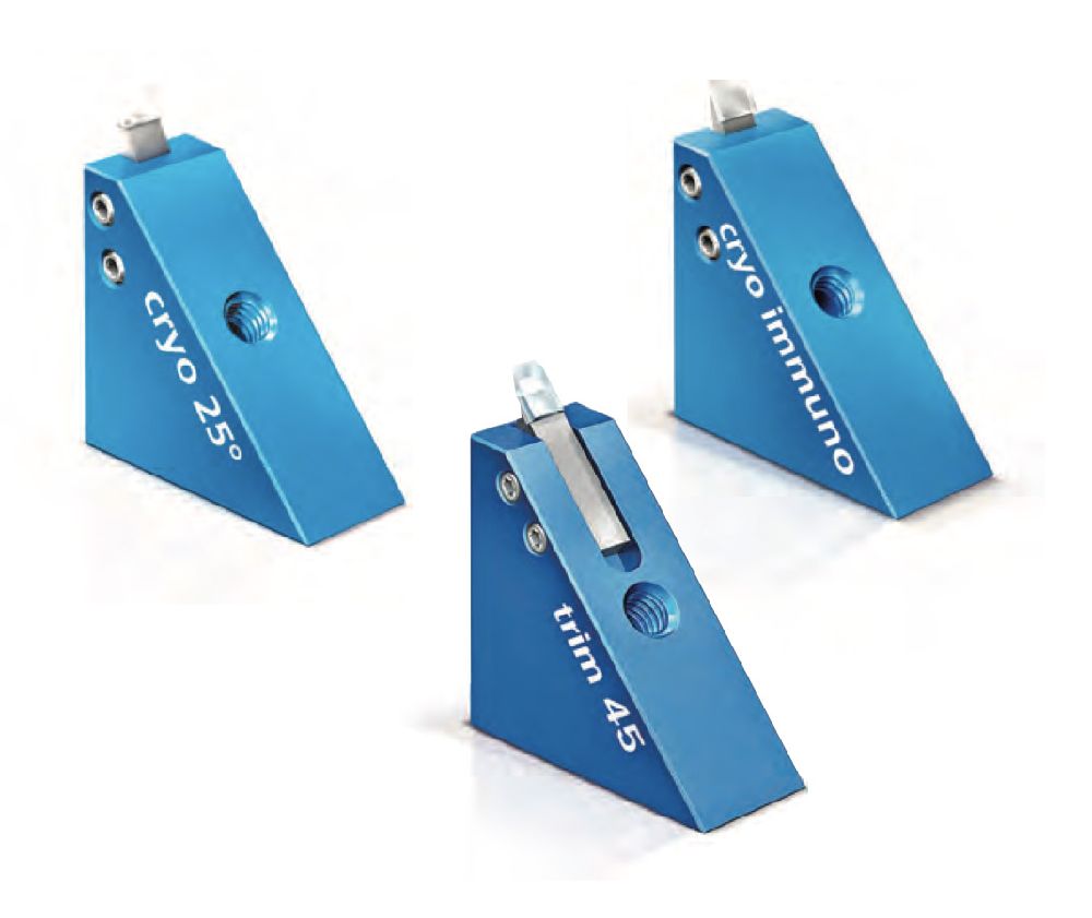
cryo 25°
The cryo 25° knife is designed for sectioning frozen hydrated specimens. The 25° angle results in the least possible compression and the best structure preservation (H.M. Han et al., Journal of Microscopy, Vol. 230, Pt. 2, pp. 167 – 171, 2007).
Please note: best results are achieved at low humidity, when the cryo-ultramicrotome is placed in a glovebox and the sections attached by electrostatic force (J. Pierson et al., Journal of Structural Biology 169, pp. 219 – 225, 2010).
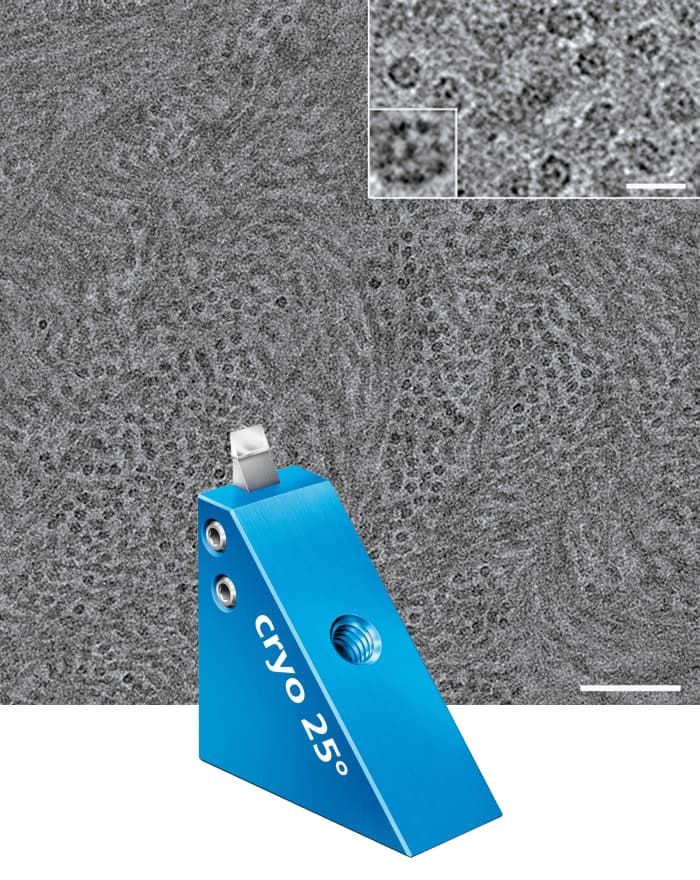
High resolution electron micrograph of vitreous section of keratin intermediate filaments in the midportion of stratum corneum of human epidermis. The fine structure of the keratin filaments is well resolved and their molecular organisation is seen in favourable cases (inset). Scale bar = 100 nm. Scale bar inset = 20 nm. Ashraf Al-Amoudi, Laboratoire d‘Analyse Ultrastructurale, Lausanne.
cryo immuno
The first cryo knife with a diamond platform, guarantees the best possible sectioning for sucrose infiltrated samples (Tokuyasu).
The diamond platform guarantees an easy and gentle section pick-up.
The sections are collected directly from the diamond surface using a loop and a sucrose/methyl-cellulose droplet (W. Liou et al., Histochemistry and Cell Biology, Vol. 106, pp. 41 – 55, 1996. P. J. Peters et al., Current Protocols in Cell Biology, pp. 4.7.1 – 4.7.18, 2006).
The 35° angle leads to a considerable reduction in mechanical stresses and therefore to improved structure preservation in sucrose infiltrated samples (E. Bos et al., Journal of Structural Biology 175, pp. 62 – 72, 2011).
We recommend the cryo immuno knife also for sectioning frozen hydrated samples (CEMOVIS). The 35° angle is a good compromise between durability and cutting performance (A. Al-Amoudi et al., Journal of Structural Biology 150, pp. 109 – 121, 2005).
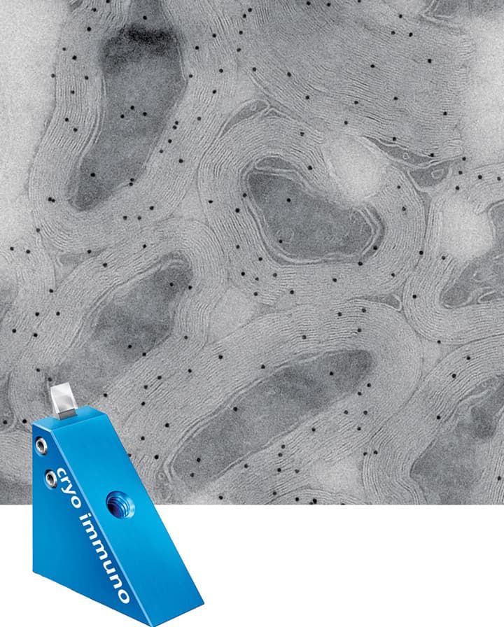
Mouse optic nerve, immunolabeling of the major myelin protein proteolipid protein (PLP), 10 nm gold. Wiebke Möbius, Dept. of Neurogenetics, EM Core Facility, MPI of Experimental Medicine, Göttingen.
cryo 35° & cryo 45° (wet/dry)
The cryo 35° knife has demonstrated its usefulness as a standard knife for the low temperature sectioning of polymers, rubber, paints, etc.
The 35° angle represents a balanced compromise between section quality and durability.
The cryo 35° and cryo 45° knife mounted in the triangular holder is suitable for dry cryosectioning.
The cryo 35° and cryo 45° knife mounted in the trough are used for sectioning with fluids such as a DMSO/water mixture.
The cryo 45° knife is well suited for routine cryo sectioning.
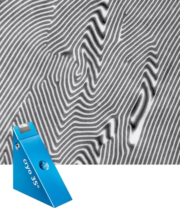
Styrene-butadiene block copolymer x 25‘000 Ronald Walter, BASF Aktiengesellschaft, Polymer Physics, D-67056 Ludwigshafen.
cryo AFM
Our cryo AFM knives are of the highest quality to ensure the increased quality requirements of AFM investigation. They produce extremely smooth sample surfaces and guarantee the best possible structure preservation.
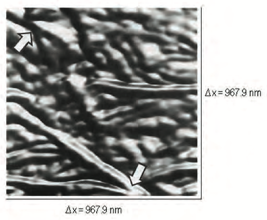
Top-view, light shaded AFM image of a cryomicrotomed surface of ultrahigh molecular weight polyethylene. The arrows indicate zones with lamellae splitting. P.H. Vallotton, Materials Science Division, Lawrence Berkley Laboratory. References Ref 1: P.H. Vallotton, M.M. Denn, B.A. Wood and M.B. Salmeron: Comparison of medical-grade ultrahigh molecular weight polyethylene microstructure by atomic force microscopy and transmission electron microscopy. J. Biomater. Sci. Polymer Edn., Vol 6, No. 7, 609-620, 1994. Top of Page
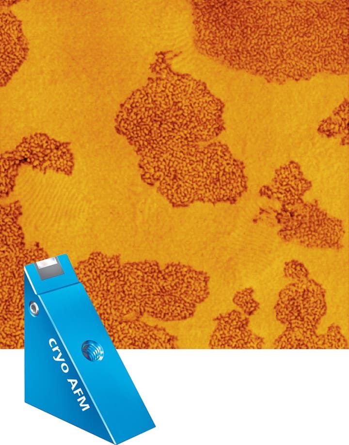
Morphology of a blend of two SBS block copolymers with different chain-architecture. AFM tapping mode, phase image, image size = 3 x 3 μm. Rameshwar Adhikari, Institut für Werkstoffwissenschaft, Martin-Luther-Universität, Halle-Wittenberg.
Diamond Knives for CEMOVIS
For the cryo Electron Microscopist, we are pleased to present the DiATOME knives for sectioning vitrified cells and tissues.
Our application specialist is at your disposal for any further information or assistance you might require.
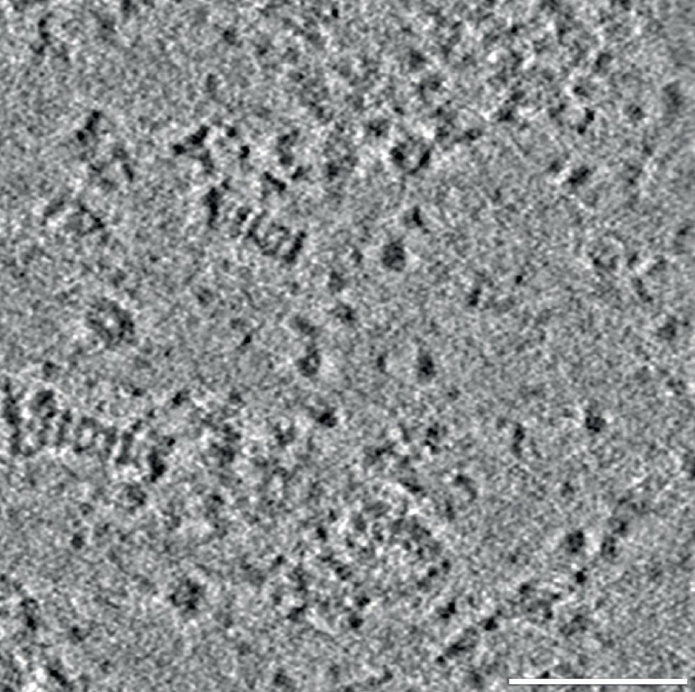
26S proteosomes within cell nucleus of a Drosophila melanogaster neuron. M. Eltsov, IGBMC, Strasbourg, Scale bar: 50nm
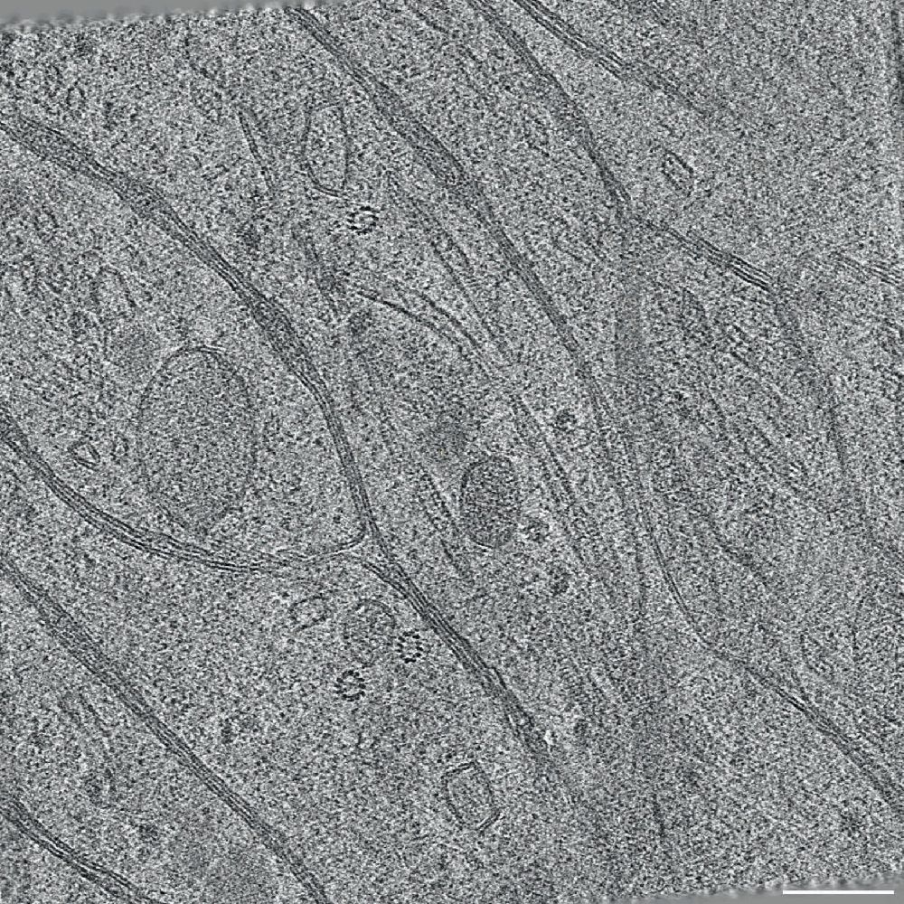
Drosophila embryonic brain axons. Defocus -4µm, no phase plate Tomographic reconstruction, computational 5nm slice. M. Eltsov, IGBMC, Strasbourg Scale bar: 100nm


